
About Thermal Imaging
 Digital Infrared Thermal Imaging (DITI) is a non-invasive diagnostic tool that shows the changes in skin surface temperature. This enables the examiner to assess physical injuries, disease and its progression, and validity of treatment by showing thermal abnormalities in the body.
There is a high degree of thermal symmetry in the body. Abnormal subtle temperature changes creating asymmetries can indicate pathology or dysfunction. DITI can help identify these asymmetries, and often at earlier stages than other diagnostics can see.
How does DITI work?
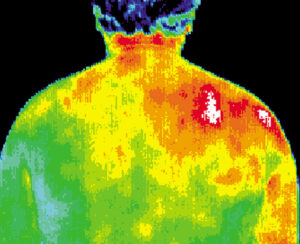
An infrared screening device is used to convert infrared radiation emitted from the skin surface into electrical impulses that are visualized into color on a monitor. The visual image graphically maps the body temperature and is referred to as a thermogram. The spectrum of colors indicates an increase or decrease in the amount of infrared radiation being emitted from the body surface. Since there is a high degree of thermal symmetry in the normal body, subtle abnormal temperature asymmetry’s can be identified.
Clinical Uses of DITI:
1. Define the extent of a lesion of which a diagnosis has previously been made
2. Localize an abnormal area not previously identified, so further diagnostic tests can be performed.
3. Detect early lesions before they are clinically evident
4. Monitor the healing process.
Skin blood flow is under the control of the sympathetic nervous system (SNS). In a healthy person, there is a symmetrical dermal (Skin) pattern which is consistent and reproducible for any individual. DITI measures the somatic component of the sympathetic nervous system to assess dermal blood flow. SNS is stimulated at the same anatomical location as its sensory counterpart and produces a somatic sympathetic response. This appears on DITI as a localized area of altered temperature with specific features for each anatomical lesion.
The pathology is generally an inflammatory process; muscle injury, epicondylitis, tendon sheath, and synovitis of joints. Both hot and cold responses may coexist if the pain associated with an inflammatory focus excites an increase in sympathetic activity. Vascular conditions are readily demonstrated by DITI including DVT, Raynaud’s disease, Vasculitis, Limb Ischemia, and others.
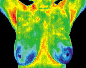
Knoxville Thermography can help you plan for better breast health by establishing a baseline on your normal thermal patterns. So if there are any changes in in your thermal patterns no matter how subtle, can be easily detected years before your self- breast exam, physician breast exam, and mammography alone. The early detection of thermal asymmetries allows you and your physician to come up with the best plan of treatment for you. It is important to state, that DITI is not a replacement for structural testing like Ultrasound or Mammography and works best in conjunction with these for the best breast health.
Using thermography as preventive medicine is not limited to just breast health. Routine baseline imaging can be used for artery disease, thyroid inflammation, disc disease, stroke, and digestive disorders.
All scans are performed by Certified Clinical Thermographers. Results and images are interpreted by Board Certified MD/OD Thermologists.